What is Basal Cell Carcinoma?

Basal cell carcinoma-BCC, is the most common skin cancer in New Zealanders.

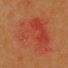
It arises from the bottom layer of the skin surface and generally grows very slowly over a number of months. It virtually never spreads to other parts of the body. It can present in a number of ways ranging from a red scaly patch of skin, to a pearly lump with prominent blood vessels on the surface. If left untreated the cancer will continue to grow and may cause ulceration of the skin.

Basal cell carcinoma is almost always caused by excessive exposure to ultraviolet light. Basal cell carcinoma, if neglected or inappropriately managed can give rise to significant morbidity and even death. There are a number of useful and proven treatment modalities, including surgical removal, Mohs Surgery, liquid nitrogen and topical creams.

It is estimated that approximately 40% of New Zealanders will develop a basal cell carcinoma in the course of their life time. Basal cell carcinoma is also the most common cancer occurring in men.

Basal cell cancers rarely spread to other parts of the body and thus rarely cause death although they can and do result in considerable inconvenience and discomfort, disfigurement and scarring.

skin specialist at skin centre

Common Questions

What is basal cell carcinoma?

Basal cell carcinoma is a type of skin cancer. It is the most common type of cancer in New Zealand, Australia and the United States. Basal cell carcinoma begins in the basal cells — a type of cell within the skin that produces new skin cells as old ones die off.It is the most common skin cancer in the New Zealand. It tends to grow slowly, locally and rarely spread distally.

What causes a basal cell carcinoma?

The commonest cause is too much exposure to ultraviolet (UV) light from the sun. BCCs can occur anywhere on your body, but are most common on areas that are exposed to the sun, such as your face, head, neck and ears. It is also possible for a BCC to develop where burns, scars or ulcers have damaged the skin. Basal cell carcinomas are not contagious.

Who is most likely to have a basal cell carcinoma?

  • People with pale skin who burn easily and rarely tan (generally with light coloured or red hair, although some may have dark hair but still have fair skin)
  • Those who have had a lot of exposure to the sun, such as people with outdoor hobbies or who work out of doors.
  • People who use sun beds or sunbathe.
  • People who have previously had a basal cell carcinoma.

If you belong to one of these groups, a regular skin check by a dermatologist is recommended to detect and treat skin cancers at an early stage.

Are basal cell carcinomas hereditary?

Apart from a rare familial condition called Gorlin's syndrome, basal cell carcinomas are not hereditary.
There is also a familial tendency, if your parents or siblings have had BCC, you may too.

What does a basal cell carcinoma look like?

BCC can vary greatly in their appearance, but people often first become aware of them as a scab that bleeds occasionally and does not heal completely. Some BCC are very superficial and look like a pinky red flat mark; others have a pearl-like rim surrounding a central crater. If left for years the latter type can eventually erode the skin causing an ulcer; Other BCC are quite lumpy, with one or more shiny nodules crossed by small but easily seen blood vessels. Occasionally the BCC can look like a scar. Most BCC are painless, although sometimes can be itchy or bleed if caught on clothes or picked up.

How will my basal cell carcinoma be diagnosed?

Sometimes the diagnosis is clear from its appearance. If further investigation is necessary to confirm the diagnosis then a small area of the abnormal skin (a biopsy) or the entire lesion (an excision biopsy) may be cut out and examined under the microscope. You will be given a local anaesthetic beforehand to numb the skin.

Can a basal cell carcinoma be cured?

Yes, basal cell carcinomas can be cured in almost every case, although treatment becomes complicated if they have been neglected for a very long time, or if they are in an awkward place - such as near the eye, nose or ear

How can a basal cell carcinoma be treated?

Surgery is the first choice of treatment.The most common surgical technique is simply to cut the BCC away, along with some clear skin around it, using local anaesthetic to numb the skin.Mohs surgeryis the treatment of choice for BCC in the head and neck area.

What is Mohs surgery? What are its advantages?

Advanced surgery, using the Mohs' technique is performed by a dermatologist with special training in the procedure.It is the treatment of choice for Non melanoma skin cancers in the head and neck area. It involves the excision of the affected skin that is then examined all the margins under the microscope straight away to see if all the BCC has been removed. If any residual BCC is left at the edge of the excision further skin is excised from that area and examined under the microscope and this process is continued until all the BCC is removed.

Mohs surgery is a tissue sparing surgery and offers the highest cure rate of any form of treatment.

What is the cure rate for basal cell carcinoma after Mohs surgery?

For primary tumour the cure rate is 99%. If the tumour is recurrent the cure rate is 95%.

What it is the cure rate for standard excision?

Analysis of large studies shows excision rate for new BCC to be 95% and recurrent BCC of 80%

What other available treatments for basal cell carcinoma?

  • Radiotherapy - shining X-rays onto the area containing the BCC. It is used as an additional treatment to surgery when required or occasionally to treat inoperable basal cell carcinomas. It has a 90% cure rate

  • For very superficial BCC:

Where the cancer cells have not invaded deeply into the skin, the following treatment options are available:

    • Cryosurgery- freezing the BCC with liquid nitrogen is done at our centre after numbing the skin with local anaesthetic. The cure rate in our department is 99%.
    • Curettage and cautery - the skin is numbed with local anaesthetic and the BCC is scraped away (curettage) and then the skin surface is sealed by heat (cautery). The cure rate is between 60 – 70%.
    • Creams – these can be applied to the skin. The two most commonly used are 5-fluorouracil (5-FU) and imiquimod. The cure rate is less than 70%
    • Photodynamic therapy – Applying a special cream to the basal cell carcinoma under a dressing for 3 hours with, which then destroys the basal cell carcinoma when a special light is shone onto it. The cure rate is around 50%

  • Blood Root "Black Salve" this natural remedy contains zinc chloride paste which indiscriminately destroys both normal and cancer cells. Typically it leave unsightly scars with no guarantee of complete treatment.

Given the lower cure rate noted with other treatment modalities, surgical excision is therefore the treatment of choice.


My doctor told me that my basal cell carcinoma is high risk. What does that mean? How would it affect me?

"High risks BCC" means it is more likely to grow back after treatment (recur).

Factors that make BCC a high risk one are:

  • Location of the basal cell at the central face, around the eyes, nose, lips and ears
  • Size of BCC > 2 cm
  • Certain BCC histological subtype (especially morphoeic, infiltrative, micronodular and basosquamous subtypes). These terms describe the organisation of the basal cell cancer cells when examined under the microscope.
  • The BCC cells invading nerves (Nerves > 0.1 mm in size)
  • Poor clinical definition of basal cell margins
  • Recurrent basal cell carcinomas.

Because recurrent basal cell carcinomas tend to have a lower response rate to treatments, it is very important to choose the most appropriate treatment modality from the outset.

Your best chance of treating a BCC and avoiding extensive scarring or disfigurement, is the first time the cancer is treated. Each time the BCC is ineffectively treated, it becomes harder to cure.

My doctor told me that my basal cell carcinoma has not been completely excised, Should I just monitor the area?

No. A further excision is always recommended. Studies have shown that 40% of the incompletely excised BCCs grow back in less than 5 years. By the time the recurrence become apparent the cancer would have spread much more extensively compared to what would have been if the cancer was treated appropriately from the beginning. Particularly with "aggressive subtype" of BCC monitoring the area is NOT a wise treatment plan because the cancer can grow extensively under the skin with little or no sign on the surface.

Self care (What can I do?)

Treatment is much easier if your SCC is detected early. BCC can vary in their appearance. You should see your doctor if you notice a lesion on your skin that is :

  • growing
  • bleeding and never completely healing
  • changing appearance in any way

Top sun safety tips:

Choose to spend time in the shade between 10am and 4pm when it's sunny. Step out of the sun before your skin has a chance to redden or burn.

  • Use sunscreen daily.
  • When choosing a sunscreen look for a high protection SPF (SPF 50 or more) to protect against UVB, and the UVA. .Apply plenty of sunscreen 15 to 30 minutes before going out in the sun, and reapply every two hours and straight after swimming and towel-drying.
  • Sunscreens should not be used as an alternative to hat , clothing and shade, rather they offer additional protection. No sunscreen will provide 100% protection.

Are there different types of Basal Cell Carcanoma?

Yes. There are. Some of the types of Basal Cell Carcinoma are -

Nodular and Superficial Basal Cell Carcinoma

These are the most common and least dangerous type of basal cell carcinoma. They are the most common variety to occur on the trunk and limbs. Principally nodular, they usually present as an early pink nodule on the skin which ultimately form a scab or bleed easily. If more superficial in variety they tend to appear as a glistening pinkish patch on the skin, the edges of which may be hard to define. Sometimes these types of basal cell carcinoma can contain pigment in which case they can be easily mistaken for melanoma.

Micronodular Basal Cell Carcinoma

This type of basal cell carcinoma occurs most commonly on the head and neck, particularly in young ladies. Often appears as a faint whitish or pearly area. There is often more cancer present than appears on the skin surface.

Infiltrative Basal Cell Carcinoma

This is also a common subtype of basal cell carcinoma to occur on the face. It typically appears as a whitish or pearly patch of skin. Once again there may be more tumour present than is indicated by the appearance. The tumour grows with widespread fine rootlets which are invisible from the skin surface. Other variants of this subtype include morphoea form or sclerosing basal cell carcinoma where the appearance may be somewhat scar-like due to the presence of increased collagen within the tumour and metaplastic or basosquamous cell carcinoma which has an appearance intermediate between that of basal cell carcinoma and squamous cell carcinoma.

Peri-neural invasion

Sometimes infiltrative types of basal cell carcinoma show a tendency to spread down nerves. This can make their removal by standard treatment methods very difficult.

In reality basal cell carcinomas often show a mixed pattern. Unsuccessful previous treatment of basal cell carcinoma tends to select a more aggressive element of the basal cell carcinoma and a tumour which first appeared nodular may after several inadequate treatments develop a substantially infiltrative growth pattern.

How quickly does Basal Cell Carcinoma grow?

Basal cell carcinoma tends to be a slow growing cancer. It tends to follow the "path of least resistance". It is for this reason that involvement of bone cartilage and muscle is not so common but tends to be a later phenomena. This may partially explain the difficulty in the management of basal cell carcinoma in areas like the eyelid, ear, nose and scalp.

In such areas, basal cell carcinomas, when they reach muscle, cartilage of bone planes, tend to spread sideways and their true extent may be masked by the appearance of normal looking overlying skin.

Get treated at the Skin Clinic

elegant reception area at skin centre

How is Basal Cell Carcinoma Treated?

It is very clear from the medical literature that treatment of basal cell carcinoma should be carried out with the intention to cure. The first time you treat a basal cell carcinoma is also the best opportunity to completely eradicate it as subsequent treatments have a lower chance of success.

The four principles that we use at the Skin Centre in the management of basal cell carcinoma are used widely by Dermatologists:

  1. Total removal or destruction of the basal cell carcinoma
  2. Preservation of normal tissue
  3. Preservation of function
  4. Optimal cosmetic result

The most important aim is to rid the patient of the tumour. If this goal is not achieved then ultimately the others will not be met. Cosmetic result should not come at the risk of failure to cure the patient.

Basal cell carcinomas grow slowly and rarely metastasize so they are sometimes not treated with the respect that they deserve. When discussing the treatment options with your dermatologist it is important to be aware of the likely recurrence rate for any given
modality of treatment for your particular type of basal cell carcinoma in its individual location.

Cryosurgery

Cryosurgery is probably one of the most common modalities used to treat basal cell carcinoma. However, in order to be effective this treatment needs to be carried out in the right manner. Ten year disease free rates of 95% are reported in the literature for
selected tumours in selected locations. Appropriate margins should be used, e.g. 4-5mm in a superficial lesion and freeze times of at least 30 seconds. This treatment in our clinic is done under local anaesthetic and the area treated usually subsequently blisters, scabs and heals over 5-6 weeks, ultimately leaving a stellate white slightly indented scar. This is the most common method we use for superficial basal cell carcinomas on the back. This is not a good treatment for recurrent basal cell carcinomas. In basal cell carcinomas greater than 1.0cm diameter or nodular lesions, cryosurgery can be combined with curettage to debulk and better delineate the size of the tumour.

Curettage and Electrodessication

This is a simple procedure practised by Dermatologists and involves scraping out the bulk of the tumour with a small sharp spoon like instrument called a curette. This is then followed by treatment of the tumour bed with electric current producing burning of the surrounding tissue to approximately 1mm in diameter. This is then repeated in 3 cycles to give an effective 3-4mm margin on the tumour. This is the best practiced for small nodular tumours, not on the face. In one study treating basal cell carcinomas on the head and neck with this modality; persistent tumour was detected in 30-40% of basal cell carcinomas, but in only 8% of those on the trunk and extremities. It is not an appropriate treatment for morphoeic or recurrent basal cell carcinomas because of poor cure rates.

Excision

This is probably the most common treatment for basal cell carcinoma. A margin of normal skin is taken around the cancer to ensure eradication of unsuspected subclinical (invisible) spread. This treatment modality offers the advantage of eventual pathological analysis providing an estimate of whether the cancer has been completely removed. One study suggested that for well defined non morphoea form basal cell carcinoma 2cm or less in diameter, 4mm margins were required to eradicate 98% of lesions.

Mohs Micrographic Surgery - What is Mohs surgery?

Mohs Micrographic Surgery offers the gold standard of treatment for basal cell carcinoma, and is well established for this in the medical literature.

Watch Video

Mohs Micrographic Surgery (see Mohs Micrographic Surgery page) offers the highest documented cure rates as well as tissue conservation of any modality for the treatment of basal cell carcinoma. It is however, a very time consuming and expensive modality. Therefore its use is reserved in our clinic for tumours on the face, ears, scalp and neck where tissue conservation is of the most importance and where strict histological control is most paramount. To achieve the high cure rates claimed Mohs Micrographic Surgery requires an extensively trained sub specialised Dermatologist and an on-site laboratory. The Dermatologist removes the tissue, colour codes it, the tissue is then mapped out and frozen sectioned on a cryostat. Microscope sections are then stained to enable identification of residual basal cell carcinoma. The slides are then examined under the microscope. Residual tumour can be identified and traced to its location in the defect using the map. The residual tumour can then be resected and checked again and so on, until all the tumour has been removed. A carefully prepared Mohs slide enables examination of 100% of the margin of the tumour against standard pathological specimen processing which allows examination of less than 0.1% of the margin of the tumour. Standard histological processing is completely adequate in the vast majority of circumstances, particularly for nodular tumours but may fail to provide an accurate indication of complete excision of recurrent tumours or primary infiltrative, morphoeic or micronodular subtypes. Mohs Micrographic Surgery provides the most effective modality also for treating tumours that show spread down nerves. Cure rates as high as 99% have been reported for primary basal cell carcinomas using this modality and as high as 97% for recurrent tumours.

The limitations of Mohs surgery include lesions with multiple foci of recurrence, tumours so invasive as to need removal of bone or deeper organs where general anaesthesia is necessary. Mohs Micrographic Surgery is almost always carried out under local anaesthesia as it is too unwieldy to provide this procedure under general anaesthesia.

Find Out More About Mohs Surgery

Radiation therapy

Radiation therapy can be an effective modality for most types of basal cell carcinomas excluding morphoeic basal cell carcinoma. It is generally not recommended for those under the age of 65 because of the long term risk of further basal cell carcinoma developing in the irradiated skin and because of the poor cosmetic results in the long term. Multiple visits are necessary for eradication of tumours, typically 15-20 visits over a 3-4 week period. For the very elderly with multiple basal cell carcinomas this can be an effective modality allowing treatment of multiple tumours in the same treatment schedule. Recurrent tumours, large tumours, tumours in high risk areas and basal cell carcinomas of the aggressive histological growth pattern are less likely to be treated well with radiation therapy. However this may be traded off against being able to treat multiple tumours at the same time without the need for reconstructive surgery.

Lasers

Carbon dioxide laser has been used most often in place of curettage and electrodesiccation as a destructive modality to treat patients with multiple basal cell carcinomas. The disadvantage of this technique, as with all destructive modalities, no specimen is available for histological analysis to determine adequate treatment of the specimen, and in addition carries the added disadvantage over curettage of being less likely to give an accurate indication of the depth of the original tumour.

Photodynamic Therapy

Photodynamic Therapy is an evolving method for the treatment of basal cell carcinoma. Its place in our practice is principally in the treatment of superficial basal cell carcinoma in the lower leg, which might otherwise have to be treated with wide excision and skin grafting. It may also be a useful treatment for superficial basal cell carcinomas in other modalities. It has a reported cure rate of 90% for superficial basal cell carcinomas. A photosensitizing cream is placed over the repaired tumour site. The patient returns in 3 hours (MAL-PTD) for irradiation of the site with red light. Swelling and sometimes crusting ensues over the following week. The lesion is then re-treated in a week's time to achieve 90% cure rates for basal cell carcinomas. Cure rates are generally less than 80% for simple nodular tumours and it is not recommended for recurrent basal cell carcinoma or basal cell carcinoma with aggressive growth pattern.

Imiquimod

Imiquimod is a relatively new treatment for basal cell carcinoma. Once again it has shown good results in studies of treatment of superficial basal cell carcinoma. Unfortunately the medical literature is confounded by the fact that the vast majority of studies for the use of Imiquimod in the treatment of superficial basal cell carcinoma were sponsored by the company making the product and further research by independent organisations is awaited. Nonetheless 90% cure rates of superficial basal cell carcinomas are reported in the literature. In our hands this is more typically around 75-80%, falling to less than 50% in treatment of tumours below the knee.

5-Fluorouracil

5-Fluorouracil is undergoing a resurgence of interest in the treatment of superficial basal cell carcinoma currently in the United States. The medical literature reports badly recurrent basal cell carcinomas following treatment with 5-Fluorouracil. The lesion should be first curetted then 5-Fluorouracil applied under occlusion. It should be applied twice daily for no less than 6 weeks, probably for 3 months. It should be reserved for patients with superficial basal cell carcinomas where no other treatment is practical.

team photo - skin centre
skin specialist at skin centre

Which treatment is right for me and my skin cancer?

Your dermatologist considers the following variables when assessing and discussing with you the best treatment for your basal cell carcinoma.

  • The size of the lesion
  • Distinctness of the tumour borders
  • Whether the tumour is primary or whether it has been treated in the past
  • Its anatomic location
  • Particular subtype of basal cell carcinoma
  • Your age and cosmetic results
  • The number of lesions you have or may have in the future


A patient who is not concerned about the cosmetic appearance may be happy with modalities such as cryosurgery or curettage and electrodesiccation. However younger individuals tend to prefer an all but invisible scar that only excision can give. The vast majority of basal cell carcinomas can be quickly excised without the need for general anaesthesia. This is particularly important in elderly patients with multiple medical risk factors. The number of lesions is important. In a younger patient presenting with more than one lesion on the face, consideration needs to be given to the possibility that they may develop many further basal cell carcinomas over the course of their life, therefore tissue conservation, cure rate and cosmesis becomes of utmost importance. This is where Mohs micrographic surgery has its advantage. The patient presenting with multiple superficial basal cell carcinomas on the back is probably best managed as far as possible with destructive modalities such as cryosurgery applied at this time in a controlled and careful fashion. A tumour which is large but also deep is better treated with wide excision and grafting or Mohs micrographic surgery. Tumours which are well defined clinically are more likely to be treated successfully with curettage and electrodesiccation or excision. Tumours which are ill defined on the face are best treated with Mohs micrographic surgery as they are most likely to be of aggressive subtypes.

It is clear from the medical literature that recurrent basal cell carcinoma, especially on the face is best treated with Mohs micrographic surgery. Curettage and electrodesiccation and cryosurgery have particularly poor cure rates when used to treat recurrent tumours (as low as 50%). Most studies report excision as having 80%, five year, disease free rates for recurrent tumours. Mohs micrographic surgery has been reported to have five year success rates of 97% for recurrent basal cell carcinoma.

As a general rule basal cell carcinomas on the trunk and limbs do not need to be treated with Mohs micrographic surgery. Because of its precise and fastidious method, Mohs micrographic surgery is preferred for areas of the face where tissue conservation is essential such as the nose, lips, eyelids and ears. It also has advantages in the management of basal cell carcinoma in areas of embryological fusion planes such as around the edges of the nose and the ears.

The subtype of basal cell carcinoma should dictate its treatment modality. Non aggressive patterns such as superficial basal cell carcinoma and nodular basal cell carcinoma can be effectively treated by many different methods. Aggressive subtypes which are more likely to have subclinical extension which may not be appreciated at the time of operation should have margin control such as Mohs micrographic surgery especially if they occur on the face, neck, ears or scalp.

Published Literature

Growth of Periocular Basal Cell Carcinoma

E. Tan1*, F.P.Y. Lin2, L.H.N. Sheck3, P.J. Salmon1 and S.G.J. Ng3

1 Skin Cancer Institute, Dermatologic Surgery Unit, Tauranga, Bay of Plenty, New Zealand

2 Department of Medicine, Waikato Hospital, Hamilton, New Zealand and Centre for Health Informatics, University of New South Wales, Sydney, Australia

3 Department of Ophthalmology, Waikato Hospital, Hamilton, New Zealand

*Corresponding author